
Figure 1. Effective inhibition of the cell signaling pathways downstream of EGFR and inhibition of HIF-1 transcriptional activity by cetuximab do not necessarily lead to successful inhibition of cell proliferation.
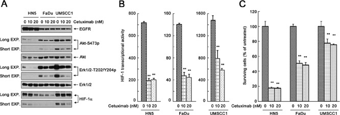
(A) HN5, FaDu, and UMSCC1 cells were cultured in 0.5% FBS medium in the absence or presence of 10 nM or 20 nM cetuximab for 24 h. Cell lysates were then prepared and subjected to Western blot analysis with the indicated primary antibodies. EXP, exposure. (B) HN5, FaDu, and UMSCC1 cells were transfected with the pBI-GL-V6L construct for 24 h. The cells were then cultured in 0.5% FBS medium in the absence or presence of 10 nM or 20 nM cetuximab in 6-well plates for 16 h. After the treatment, cell lysates were prepared for HIF-1 luciferase reporter assay. Arbitrary luciferase activity units were normalized to the amount of protein in each sample. Data shown are means and SDs (n = 3). P values for the comparisons were determined by Student's t-test. **p < 0.01. (C) HN5, FaDu, and UMSCC1 cells were cultured in 0.5% FBS medium in the absence or presence of 10 nM or 20 nM cetuximab for 5 days. The relative number of surviving cells was determined by MTT assay. The OD values of the treated groups were normalized to the OD value of untreated cells, which was set as 100%. Data shown are means and SDs (n = 3). **p < 0.01.