
Figure 2. Reduced expression of BCL6B was associated with promoter region hypermethylation in human primary HCC.
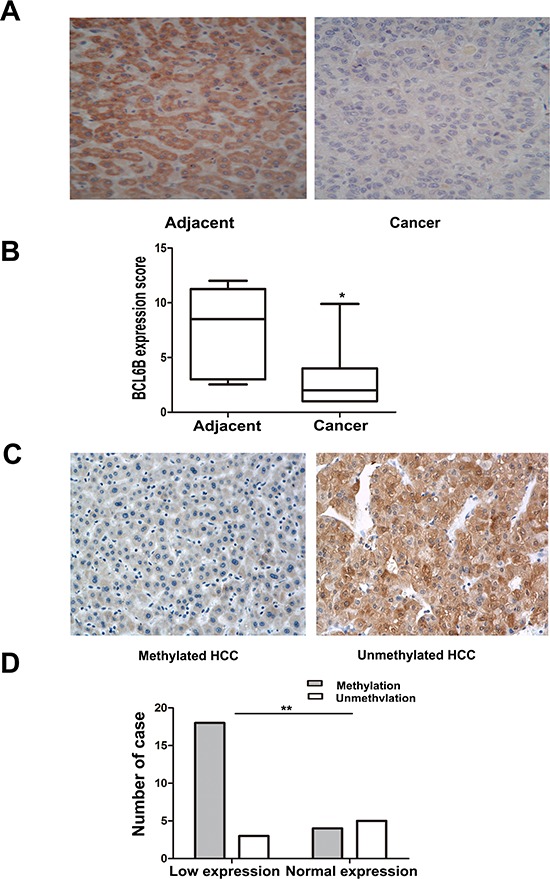
A. Representative BCL6B staining in matched human primary HCC (right) and adjacent tissue samples (left) by IHC. (X200). B. The scores of BCL6B expression in 30 matched HCC and adjacent tissue samples are shown as box plots. Horizontal lines in the boxes represent the median score; the bottom and top lines of the boxes represent the 25th and 75th percentiles, respectively; vertical bars represent the range of data. (*p < 0.05, **p < 0.01) C. Representative BCL6B staining in methylated (left) and unmethylated HCC (right) by IHC. (X200) D. Loss of/reduced expression of BCL6B was associated with promoter region hypermethylation in human HCC. (*p < 0.05, **p < 0.01)