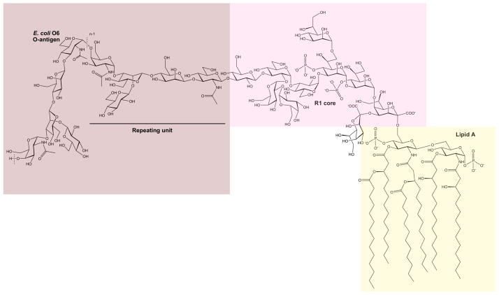
Figure 1.
Schematic of the lipopolysaccharide (LPS) of E. coli O6 having an R1 core. The LPS consists of three regions, viz., the lipid A, the core, and the O-antigen polysaccharide built of pentasaccharide repeating units. Note that the N-acetyl-D-glucosamine residue of the first repeating unit is ligated to the core via a β-linkage, whereas in the remaining part of the polysaccharide, it is joined via α-linkages to the subsequent repeating unit. The dashed lines indicate the repeating unit of the polymer and n describes the total number of repeating units in the O-antigen polysaccharide.

