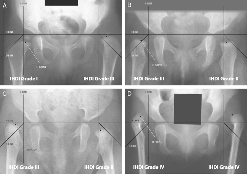
FIGURE 4.

(A) Case example of an anteroposterior (AP) pelvic radiograph of an infant with DDH. The right hip was classified as an IHDI grade I hip, whereas the left hip was classified as grade III. (B) Case example of an AP pelvic radiograph of another infant with bilateral DDH. The right hip was classified as an IHDI grade III hip, whereas the left hip was classified as grade II. (C) Third case example of an AP pelvic radiograph of an infant with DDH. The right hip was classified as an IHDI grade III hip, whereas the left hip was classified as grade II. (D) Final case example of an AP pelvic radiograph of an infant with bilateral DDH. Both hips were classified as IHDI grade IV. Note that all hips in the case examples could be classified, although the ossific nucleus was variably present.
