
Fig. 6.
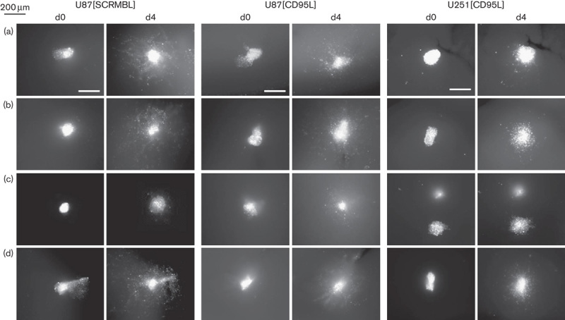
Glioma cell migration in organotypic murine brain slices. Spheroids from DiI-labelled U87[SCRMBL] (left panel), U87[CD95L] (middle panel) and U251[CD95L] cells (right panel) were inoculated into organotypic C57BL/6 mouse brain slices pretreated with (a) control medium or pretreated with medium containing (b) APG293 (10 ng/ml), (c) APG293 (10 ng/ml)+APG101 (50 μg/ml) or (d) APG293 (10 ng/ml)+APG122 (50 μg/ml). Migration of tumour cells was monitored by fluorescence microscopy at the time points indicated. Scale bar=200 μm.