
Figure 3. Etomidate evoked a greater enhancement of steady-state currents than that of peak currents.
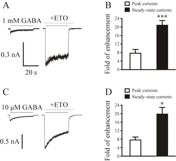
A, Representative current traces evoked by prolonged application (30 s) of 1 mM GABA as well as 1 mM GABA and 3.2 μM etomidate (with etomidate pre-applied) to show the steady-state current change evoked by etomidate for β3-α1-δ/β3-α1 receptors. The solid lines indicated the application of GABA, and the dashed lines denoted the application of etomidate. B, The mean fold of peak current and steady-state current enhancement by etomidate in the presence of 1 mM GABA for β3-α1-δ/β3-α1 receptors. C, Representative current traces evoked by prolonged application (30 s) of 10 µM GABA as well as 10 µM GABA and 3.2 µM etomidate (with etomidate pre-applied) to show the steady-state current change evoked by etomidate for β3-α1-δ/β3-α1 receptors. D, The mean fold of peak current and steady-state current enhancement by etomidate in the presence of 10 µM GABA for β3-α1-δ/β3-α1 receptors. *, Significantly different from peak current enhancement at p < 0.05; *** p < 0.001.