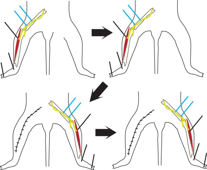
Fig. 5.
Dorsal drawings of a rat show the nerve conduction setup for stimulation studies. Stimulation is at two locations on the peroneal nerve (indicated in blue). Each animal was tested on the left leg first (above, left and above, right). The left leg was then sutured closed and the right leg exposed (below, left and below, right). Between these steps, there was a 30-minute resting period, and the electrodes were removed. Stimulating electrodes for each rat consisted of one plain and one polymerized with PEDOT. The type of stimulating electrode used on the left side was randomly assigned.

