
Figure 1.
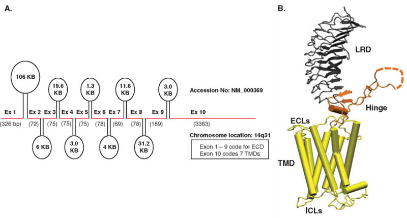
A. Genetic organization of the human TSHR receptor. The TSHR gene located on chromosome 14q31 consists of 10 exons as indicated here by the thick red line segments. The introns between these exons are indicated as ovals with their sizes marked within them. The first 9 exons of the TSHR gene code for the extracellular domain (ECD) and exon 10 codes for the transmembrane domain (TMD) of the receptor. B. Homology model of the TSH holoreceptor. The model highlights the tripartite structure of the TSHR. The ectodomain shown in gray is made up of 10 leucine-rich repeat domains (LRD with loops and β-pleated sheets obtained from the published crystal structure [9] (PDB: 3G04). The region connecting the LRD and TMD, known as the ‘hinge’ region has recently been crystallized for the FSH receptor and is shown as a looped structure (orange) with a helix conformation close to the carboxyl end of the LRD. The hinge in the TSHR has an additional sequence insert and is larger than in the FSH receptor. Therefore, amino acids 305 – 381 are missing in the illustrated model, and this insert is depicted as a closed dotted loop. The TMD (yellow), with its 7 helices, is depicted as cylindrical structures connected to each other by the specific TSHR intra- and extracellular loops (ICLs and ECLs). The TMD is the region that harbors the allosteric binding pockets for the SMLs.
A. Reproduced with permission from [95].
B. Reproduced with permission from [72].
ECL: Extracellular loops; ICL: Intracellular loops; SMLs: Small molecule ligands; TSHR: Thyroid-stimulating hormone receptor.