
Fig. 2.
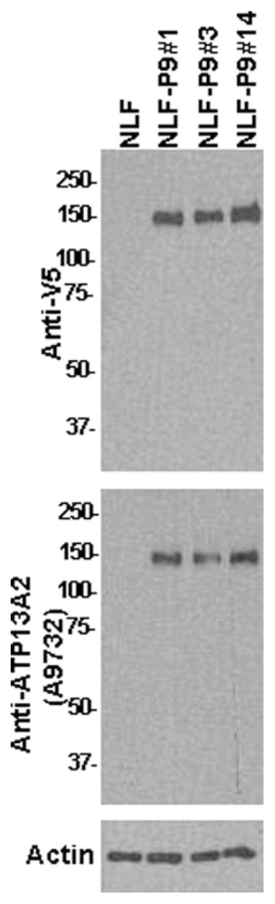
Western blot analysis demonstrating the expression of ATP13A2 in stable NLF cells clones. Western blot analysis with anti-V5 antibody or anti-ATP13A2 antibody A9732 showing the expression of WT ATP13A2 in three stable NLF cell lines (clones NLF-P9 No. 1, NLF-P9 No. 3, and NLF-P9 No. 14). One lane was loaded with protein extract from nontransfected cells. Ten micrograms of total cell extract lysed in 3% SDS was loaded in each lane of 8% polyacrylamide gels. Membranes were probed with either anti-V5 antibody or anti-ATP13A2 antibody A9732. The mobility of molecular mass markers is indicated at left. An immunoblot with an antiactin antibody is shown as a loading control.