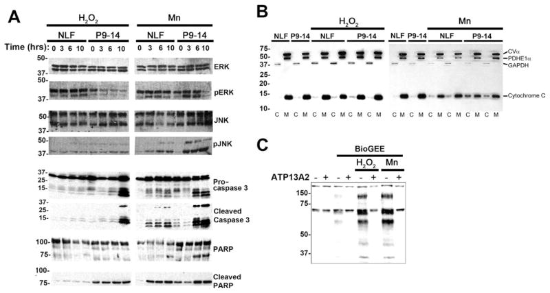
Fig. 8.
Analysis of the effects of ATP13A2 expression on signal transduction pathways under conditions of cellular stress. A: Native NLF cells and NLF P9–14 cells were challenged with 50 μM H2O2 or 1 mM MnCl2 for 0, 3, 6, or 10 hr. Western blot analysis of various components of signal transduction pathways were performed with the antibodies indicated. Equal protein amounts were loaded in each lane of SDS-polyacrylamide gels. B: Biochemical assay for the release of cytochrome C in the cytosol. Cells were treated with 50 μM H2O2 or 1 mM MnCl2 for 6 hr. C, cytosolic fraction; M, mitochondrial fraction; CVα, complex Vα; PDHE1α, pyruvate dehydrogenase E1α; GAPHD, glyceraldehyde 3-phosphate dehydrogenase. C: Native NLF cells (−) and cells overexpressing ATP13A2 (NLF P9–14; +) were assayed for intracellular oxidative damage by glutathione incorporation with the BioGEE assay. Cells were untreated or treated with 50 μM H2O2 or 1 mM MnCl2 for 6 hr.

