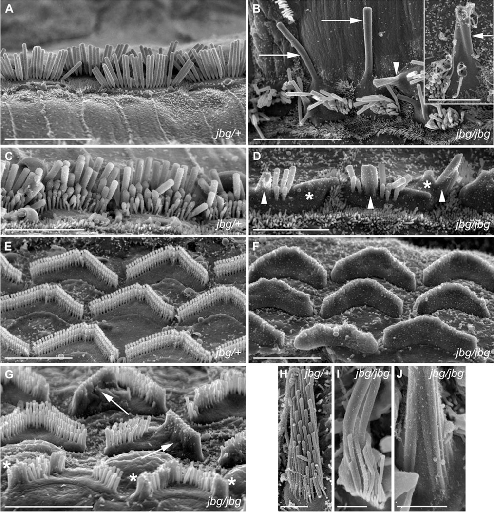
Fig. 1.
Morphological defects of hair cells in jitterbug mutant. Auditory (A–G) and vestibular (H–J) hair cells from P17 and P10 (inset) mice. (A) Inner hair cells (IHC) from a heterozygous control (jbg/+) viewed from lateral aspect near the apex of the cochlear duct. (B) IHC near the apex of a cochlea from a jitterbug mutant (jbg/jbg) showing elongated and fused stereocilia lying parallel to the surface of the epithelium (arrows), and fused stereocilia with lifted membranes (arrowhead). Inset shows IHC with fused stereocilia (arrow) at P10. (C) Control IHC at the base with staircase arrangement of stereocilia. (D) Mutant IHC at the base lack the shortest row of stereocilia and exhibit fusion of several stereocilia (arrowheads) as well as mass fusion indicated by the lifted plasma membrane (*). (E) Outer hair cells (OHC) at the base of control cochlear duct showing regular arrangement of stereocilia arrays. (F) OHC at the base of a mutant cochlear duct devoid of individual, non-fused stereocilia. (G) OHC from mid-turn of mutant cochlear duct with varying degrees of stereocilia fusion, including entire arm of V-shaped bundle (arrows) and fusion at the ends of bundles (*). (H) Utricular hair cells (UHC) from heterozygous control showing stereocilia arranged in rows of increasing height and uniform diameter within each row. (I) Mutant UHC bundle lacks a regular staircase array and displays thickened stereocilia. (J) High magnification view of proximal end of mutant UHC bundle with a morphology reflecting apparent fusion of multiple parallel actin bundles covered by a common membrane. Scale bars: 10 µm (A, B), 5 µm (inset, C, D, E, F, G), 2 µm (H–J).

