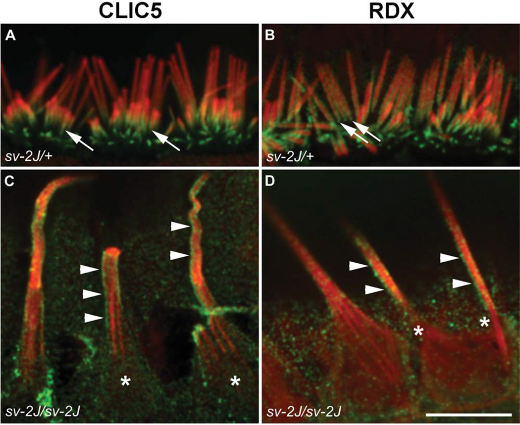
Fig. 11.
Localization of CLIC5 and RDX in hair cells from Myo6 mutant Snell’s waltzer 2 Jackson (sv-2J) mice. Inner hair cells from the apex of P15 cochleae from heterozygous control (sv-2J/+) mice (A, B) and homozygous mutant (sv-2J/sv-2J) mice (C, D). Hair cells were stained with antibodies (green) against CLIC5 (A, C) or RDX (B, D) and counterstained with phalloidin (red). Both proteins are enriched at the base of stereocilia in control cells (arrows). However, in Myo6 mutant cells, both proteins exhibit diffused staining along the shaft of giant fused stereocilia. Staining of CLIC5 and RDX is enriched at the surface of shafts (arrowheads), indicating preferential association with the membrane; accumulation of CLIC5 or RDX is no longer seen at the bases of fused stereocilia (asterisks). Scale bar: 10 µm.

