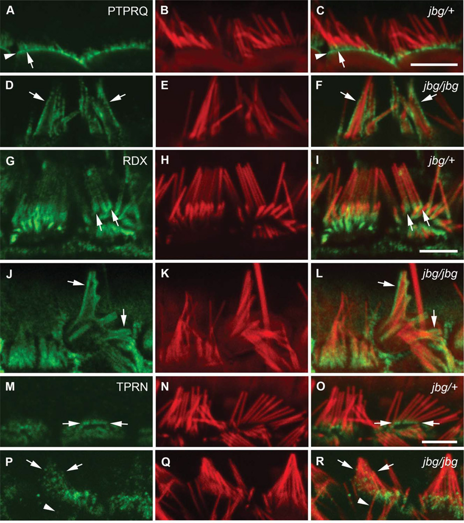
Fig. 6.
Localization of PTPRQ, RDX, and TPRN in mature hair cells of jitterbug mutant. Inner hair cells near the apex of the cochlea stained with antibodies (green) against PTPRQ at P40 (A–F), RDX at P17 (G–L), and TPRN at P21 (M–R) and counterstained with phalloidin (red). (A-F) In control cells (jbg/+), intense PTPRQ staining is restricted to the bases of stereocilia (A, C, arrow) as well as the intervening apical plasma membrane (A, C, arrowhead); in mutant cells (jbg/jbg), PTPRQ is distributed in a diffused pattern extending along the membrane covering the shaft of the still recognizable stereocilia (D, F, arrows). (G-L) Control cells exhibit enriched RDX staining at the base and a lower level of staining along the stereocilia shaft towards the tip (G, I, arrows); mutant cells exhibit a diffuse pattern of RDX staining extending along the membrane covering the shaft of the still recognizable, but severely malformed stereocilia (J, L, arrows). (M–R) In control cells, TPRN staining is enriched at the base of stereocilia (M, O, arrows); in mutant cells, TPRN staining is distributed in a diffused pattern extending along the shaft of the malformed stereocilia (P, R, arrows). TPRN staining is also dispersed along the shaft of a single individual stereocilium (P, R, arrowhead). Scale bars: 5 µm.

