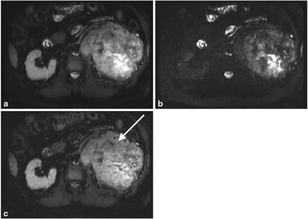
Fig. 5.
Shorter TE image (a), longer TE image (b) and the subtracted iSTIR image (c) of a patient with a large left renal mass (arrow). Note the heterogeneity of the tumor with fluid accumulation centrally that is partially suppressed in the subtracted iSTIR image. The final diagnosis of the tumor is primary clear cell renal cell carcinoma, Fuhrman grade 4 with extensive sarcomatoid features

