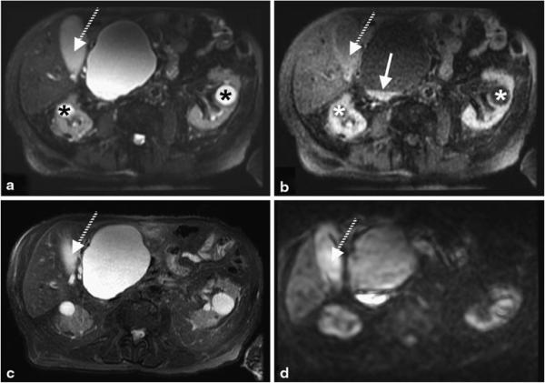
Fig. 7.
Shorter TE image (a) and subtracted iSTIR image (b) compared against fat-suppressed T2-weighted image (c) and diffusion weighted image (b = 850 s/mm2) (d). The cysts in both kidneys (asterisk) are completely suppressed and the gallbladder (dashed arrow) is largely suppressed on the subtracted iSTIR image. Note the increased signal at the bottom of the large pancreatic cyst next to the liver (arrow) on the subtracted iSTIR image, which also appears bright on the diffusion-weighted image. The final diagnosis is pancreatic pseudocyst and common bile duct stricture with biliary dilatation

