Fig. 1.

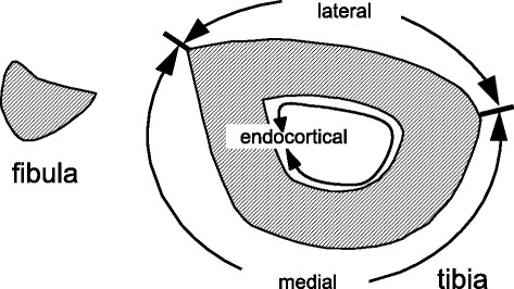
Three surfaces of the tibia for histomorphometric analysis. The tibial periosteal surface was divided into lateral and medial regions as indicated for histomorphometric analysis

Three surfaces of the tibia for histomorphometric analysis. The tibial periosteal surface was divided into lateral and medial regions as indicated for histomorphometric analysis