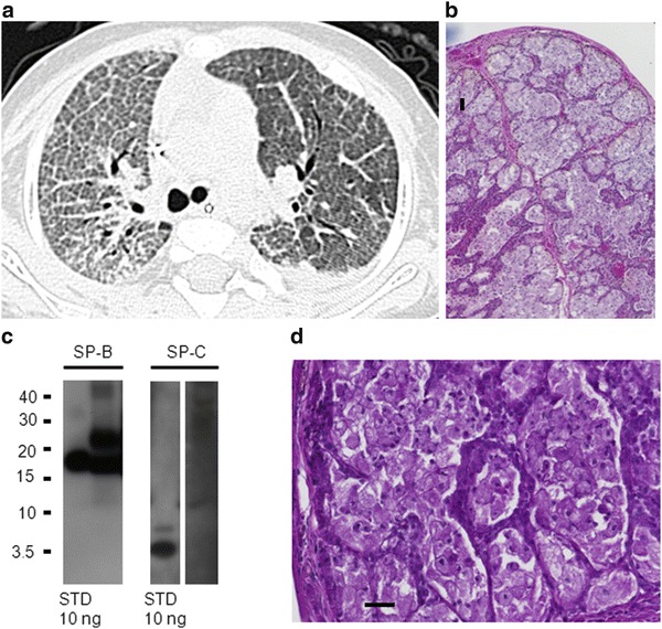
Fig. 1.

Patient 1 at the age of 7 months: (a) Upper left: CT scan showing crazy-paving pattern with pronounced inter- and intralobar septi, particular on the right side; left side after therapeutic lavage. (b) Upper right and (d) lower right: Lung biopsy (hematoxylin and eosin stain, 40-fold) showing alveolar filling with fine granular material. PAS-positive material (100 fold) mainly foamy macrophages and some extracellular surfactant material. (c) Lower left: Western blotting of bronchoalveolar lavage for surfactant protein B (SP-B) and SP-C. 5 μg of total protein of lavage fluid per lane was added and the respective standards (STD). After SDS-PAGE and transfer, the membranes were probed with antibodies against SP-B and SP-C. Molecular weights (kDa) are indicated on the left side. All bands were analyzed under nonreducing conditions. SP-B was detected as dimers (typical bands at 16 kDa; compare to standard STD of 10 ng applied to lane 1) and some higher molecular forms (bands at 24 kDa); such forms are of interest, as often seen in patients with alveolar proteinosis. No monomers or degradation products were detected. SP-C was absent at molecular weight of about 4 kDa (usually SP-C is present in amounts at least about 50% of SP-B)
