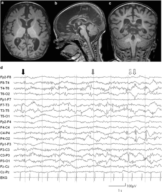
Fig. 2.

(a–c) Axial sagittal and coronal T1-weighted cranial MRI slices of patient 1, showing delayed myelinization sparing the frontal and posterior subcortical white matter and global brain atrophy; (d) EEG performed with current application of high doses of midazolam showing multiregional epileptiform discharges temporal left (black arrow), right (gray arrow), parietal left (lined arrow), and right (bare arrow). In addition, excessive beta-activity reflecting benzodiazepine effect is evident
