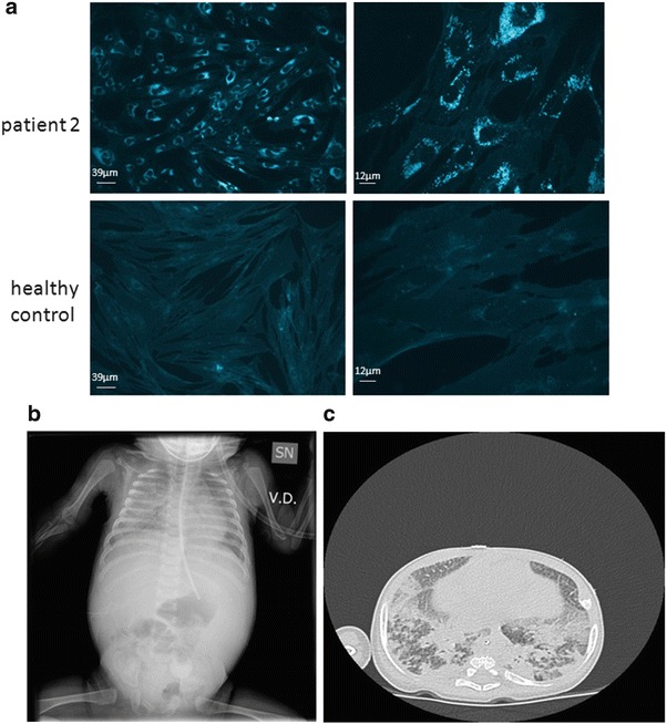
Fig. 3.

(a) Filipin staining of cultured fibroblasts in patient 2, showing a massive intracellular accumulation of unesterified cholesterol. (b, c) Chest X-ray and high-resolution CT of patient 2, showing diffuse hypo-diaphaneity with air bronchogram in the whole right pulmonary field and in the left inferior field (b) and smooth septal thickening and ground glass opacities in intermixed pattern (c)
