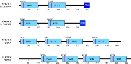
Fig. 4.

Schematic representation of human NHERF isoforms. PDZ domains are indicated by the light blue rectangular boxes and EBDs by dark blue boxes. The amino acid residues that constitute the core binding-motif are noted and shown as a purple bar indicating their location within the respective PDZ domain.
