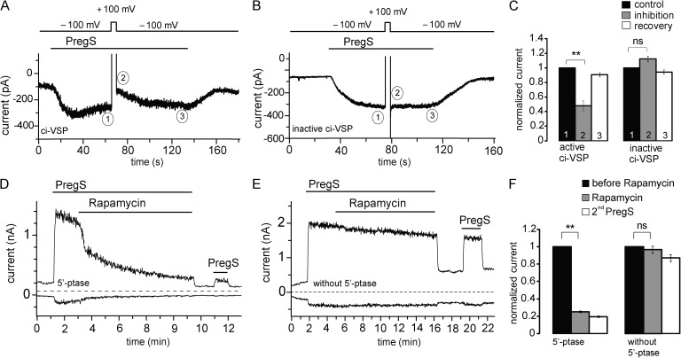
Figure 6.
Rapidly inducible 5′-phosphatases inhibit mTRPM3α2. (A) Representative trace of mTRPM3 current recorded from a HEK cell transfected with the active ci-VSP at a holding potential of −100 mV followed by short depolarizing pulse of 100 mV to activate the phosphatase. (B) Representative trace from a HEK cell transfected with the phosphatase-inactive mutant of ci-VSP (C363S) using the same voltage protocol as in A. (C) Summary of the inhibition of PregS-induced TRPM3 current plotted by comparing the current at −100 mV before and immediately after the depolarization pulse for the active (n = 6) and inactive phosphatases (n = 5). (D) Representative measurement in a HEK cell expressing the mTRPM3 and the components of the rapamycin-inducible 5-phosphatase. Measurements were performed using a ramp protocol from −100 to 100 mV, and current amplitudes are plotted at 100 and −100 mV. The applications of 25 µM PregS and 100 nM rapamycin are indicated by the horizontal lines. (E) Similar experiment as in D in a control cell, expressing TRPM3 and the components of the rapamycin-inducible system without the 5-phosphatase. (F) Statistical summary of the data (n = 6–7). Error bars represent SEM. **, P < 0.01.

