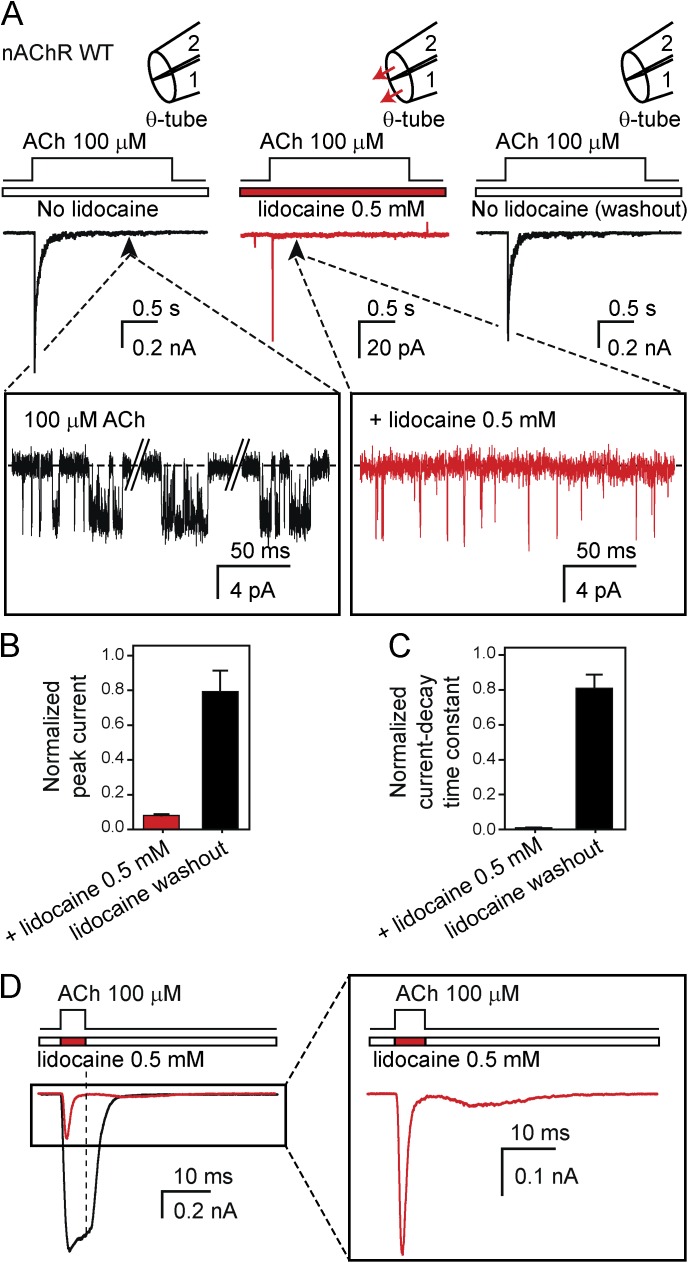
Figure 3.
Block of the muscle nAChR by lidocaine. (A) Macroscopic current responses to 2-s pulses of 100 µM ACh recorded at –80 mV from the mouse muscle adult-type nAChR in the outside-out configuration. The response of each patch of membrane was recorded sequentially in the absence of lidocaine (left), in the presence of 0.5 mM lidocaine flowing through both barrels of the theta-type perfusion tubing (middle), and back again, without lidocaine in either barrel (right). The insets show the much lower open probability of the channel in the presence of 0.5 mM external lidocaine; openings are downward deflections. The solutions flowing through the two barrels of the perfusion tubing were (in mM) 142 KCl, 5.4 NaCl, 1.8 CaCl2, 1.7 MgCl2, and 10 HEPES/KOH, pH 7.4 with or without ACh and with or without lidocaine. In the schematic representations of the theta-tubing perfusion, arrows indicate the application of lidocaine. Note the expanded current scale used for the middle panel; the trace is the average of 25 consecutive responses recorded from a representative patch. (B) Peak-current amplitudes recorded in response to the application of ACh in the presence of external 0.5 mM lidocaine and upon lidocaine removal normalized to the peak value observed in the initial lidocaine-free recording. (C) Current-decay time constants during the application of ACh in the presence of external 0.5 mM lidocaine and upon lidocaine removal normalized to the time constant fitted to the initial lidocaine-free recording. The values plotted in B and C are means obtained from 4 patches; error bars are the corresponding standard errors. (D) Comparison of the responses of the nAChR to 5-ms pulses of ACh alone and ACh plus lidocaine applied sequentially to the same patch of membrane. The vertical dashed line emphasizes the different current values observed upon fast washout.

