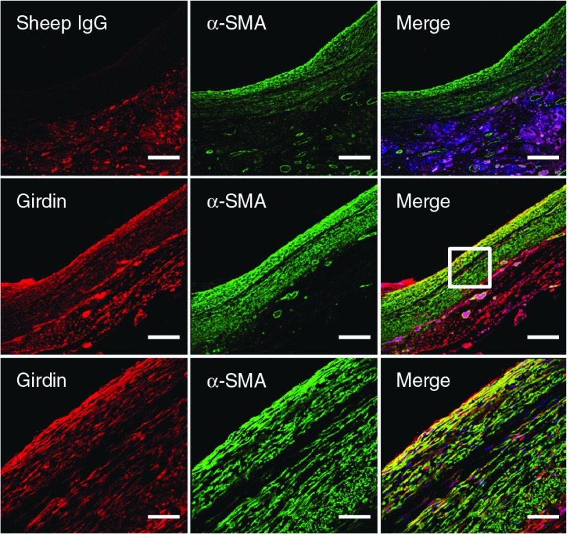
Fig. 1.
Vein grafts at 14 days post-operation were subjected to immunofluorescence staining using sheep IgG as a negative control or anti-Girdin antibody (red), and anti-α-SMA antibody (green). Cell nuclei were labeled with DAPI. Representative photos at low magnifications (upper and middle panel) are presented. The boxed area is magnified (lower panel). Bars: 200 µm (upper and middle); 50 µm (lower). α-SMA: α-smooth muscle actin

