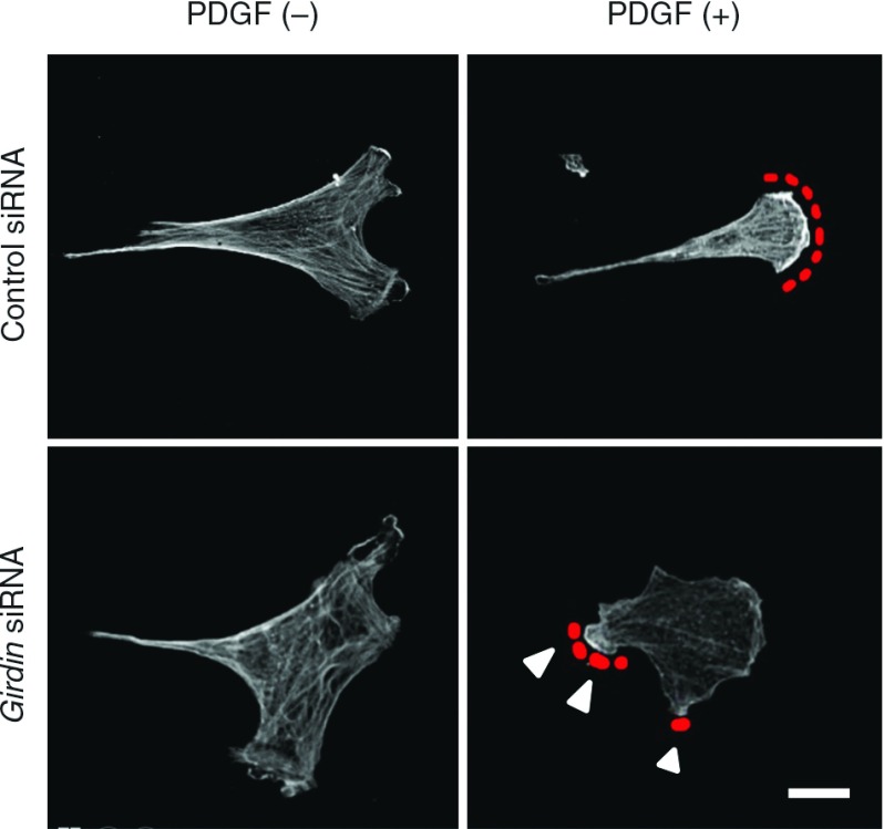
Fig. 3.
Venous SMCs were stimulated with PDGF-BB (20 ng/ml) for 10 minutes and stained with anti-β-actin antibody. Red dotted lines denote lamellipodia at the leading edge. Arrowheads denote lesspolarized and less extended membrane protrusions comparedwith the control cells. Bar: 20 µm. SMCs: smooth muscle cells

