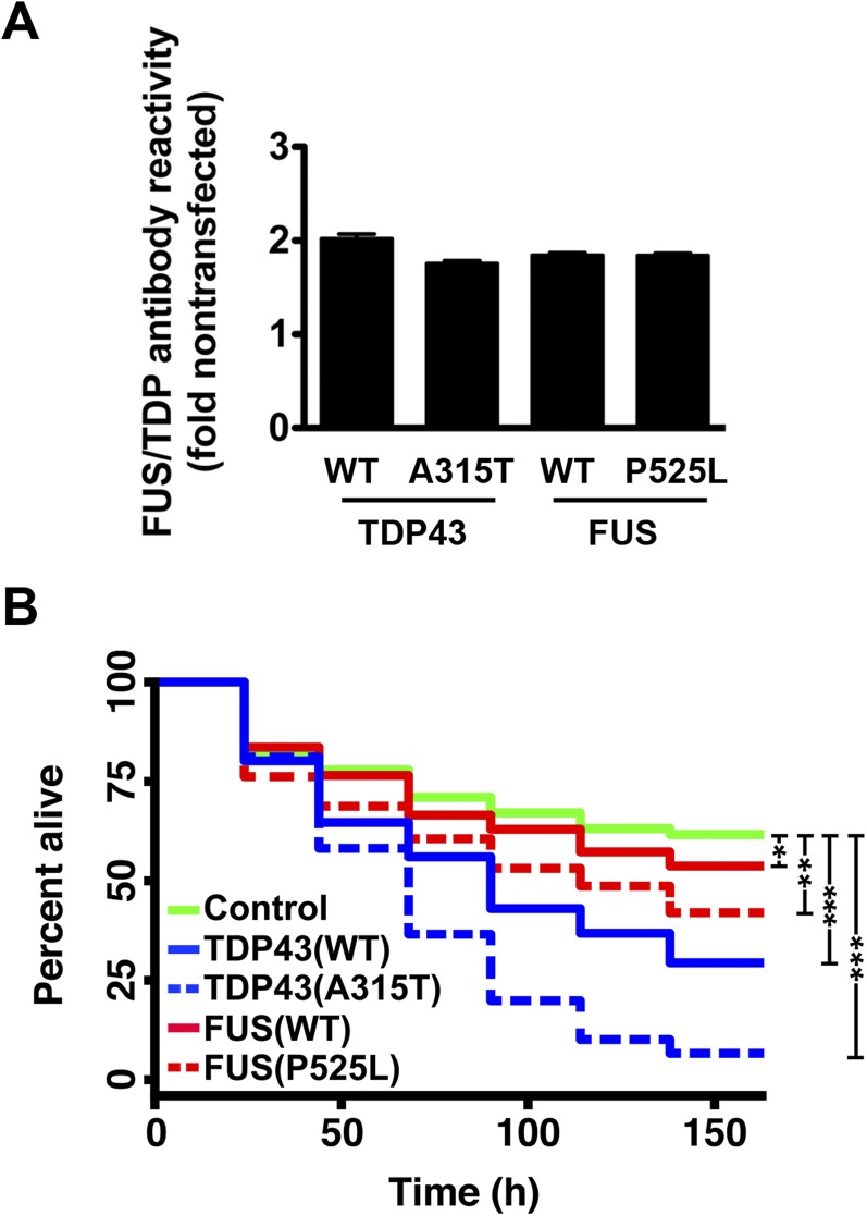
Fig. S1.
A primary neuron model of ALS. Primary rodent cortical neurons were dissected then transfected with plasmids encoding mApple or mApple-tagged TDP43 or FUS variants on day 4 in vitro. (A) Neurons transfected with TDP43 or FUS displayed an approximate 2-fold mean increase in antibody reactivity, as measured by quantitative fluorescence microscopy. n = 316–433 neurons per genotype. Error bars, ± SEM. (B) Survival of transfected neurons was followed by LFM as described in the text, and survival was plotted using Kaplan–Meier analysis. *P < 0.05, **P < 0.0001, ***P < 1 × 10⁻¹⁰, by Cox hazards analysis. n = 98–139 neurons per genotype, pooled from 8 wells each.

