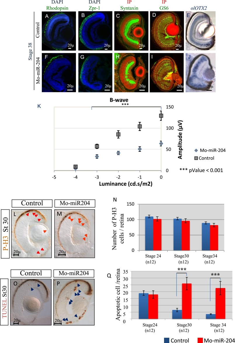
Fig. S6.
Loss of function of miR-204 has a deleterious effect on photoreceptor maintenance and function in vivo. Representative frontal eye sections of St38 control-injected (A and B) and Mo-miR-204–injected (F and G) medaka fish embryos immunostained with anti-Rhodopsin and Zpr-1 antibodies (green). Sections are counterstained with DAPI (blue). We observed a significant reduction in the intensity of both rhodopsin and Zpr-1 staining in Mo-miR-204–injected medaka fish compared with controls. Representative frontal eye sections of St38 control-injected (C and D) and Mo-miR-204–injected (H and I) medaka fish embryos immunostained with anti-Syntaxin and GS6 antibodies (green). No significant differences in GS6 staining were detected between morphants and control embryos. In Mo-miR-204–injected embryos, amacrine and ganglion cells do not form a proper inner plexiform layer, as described in ref. 22) (green signal). Sections are counterstained with propidium iodide (IP; red signal). (E and J) Frontal vibratome sections of wt and Mo-miR-204–injected medaka embryos processed for whole-mount RNA ISH with antisense probe for olOtx2. No apparent alterations in bipolar cells were observed, as determined by expression analysis of olOtx2 (E and J). (K) Intensity response curves of mean b-wave amplitude (±SEM) of control-injected and Mo-miR-204 injected larvae (n = 30). The b-wave amplitudes of morphant larvae are significantly smaller with respect to controls over the 5 units of luminance intensity tested (shown on the x axis in a logarithmic scale). (L and M) Frontal vibratome sections of St30 control-injected and Mo-miR-204–injected embryos immunostained with antiphospho-histone H3 (P-H3) antibodies. (N) Quantification of PH3-positive cells in the retina of the control-injected or Mo-miR-204–injected embryos at different embryonic stages as indicated. No significant changes were detected between morphants and controls. Red arrows, P-H3-positive cells. (O and P) Frontal vibratome sections of St30 TUNEL-stained control and miR-204 morphant embryos. (O and P) A significant increase of cell death in miR-204 morphant eyes was observed compared with control. (Q) Quantification of TUNEL-positive cells in the retina of control-injected or Mo-miR-204–injected embryos at different embryonic stages; blue arrows, TUNEL-positive cells. ***P < 0.0001 in in likelihood ratio tests. (Scale bars, 20 μm.)

