Fig. 4.
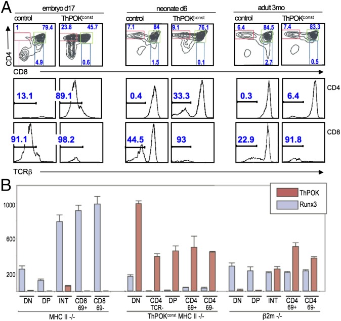
Altered early thymocyte development in ThPOKconst mice. (A) FACS analysis of thymocytes from ThPOKconst and control non-Tg mice at different stages in ontogeny, as indicated, showing expression of CD4, CD8, and TCRβ. Note prominent TCRβ− SP CD4 subset in embryonic ThPOKconst mice, which diminishes progressively in neonates and adults. Also, note that the CD8+ immature single positive (ISP) subset is absent in ThPOKconst mice at e17 (0.6% SP CD8 cells, compared with 4.9% in WT control). (B) RT-PCR analysis of ThPOK and Runx3 mRNA expression in indicated sorted thymic subsets from ThPOKconst MHC class II−/− mice, compared with MHC class II−/− and β2m−/− mice. Note that ThPOK is expressed constitutively in all subsets from ThPOKconst MHC class II−/− mice, whereas Runx3 is severely repressed in these mice. INT refers to CD4+8low subset intermediate between DP and SP stages.

