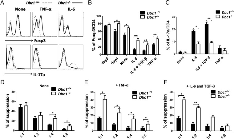
Fig. 2.
Foxp3 from Dbc1−/− mice is more stable than Foxp3 from WT mice, and Treg cells from Dbc1−/− mice are more suppressive than Treg cells from WT mice, especially during inflammatory insult. (A) Treg cells isolated from thymi of C57BL/6 Foxp3-GFP mice were expanded for 4 d and were recultured in the presence of APCs with soluble anti-CD3 (1 μg/mL), anti-CD28 (1 μg/mL), anti–IL-4 (5 μg/mL), and anti–IFN-γ (5 μg/mL), with or without recombinant human (rh)-TGF-β (5 ng/mL), recombinant mouse (rm)-IL-6 (10 ng/mL), or rm-TNF-α (50 ng/mL). After 3 d these cells were harvested and stained for intracellular expression of Foxp3 and IL-17a. (B) The statistical percentages of Foxp3 expression among the different groups as indicated. (C) The statistical percentages of IL-17a expression among the different groups as indicated. “None” indicates no rm-TNF-α or rm-IL-6 treatment. (D–F) Suppressive activity of Treg cells from Dbc1−/− and Dbc1+/+ mice. Treg cells from Dbc1−/− and Dbc1+/+ mice without treatment (“None”) (D), pretreated with 50 ng/mL rm-TNF-α (E), or treated with 10 ng/mL rm-IL-6 and 5 ng/mL rh-TGF-β (F) were tested using an in vitro suppressive activity by indirectly measuring the proliferative rates of anti-CD3–activated T-responder cells (B6) labeled with CFSE. Cells were harvested on day 3 and stained with PE-conjugated anti-CD8 antibody. Samples were analyzed by flow cytometry and gated on the CD8+ population. The x axis shows the ratio of Treg cells to responder T cells. All data are representative of three independent experiments.

