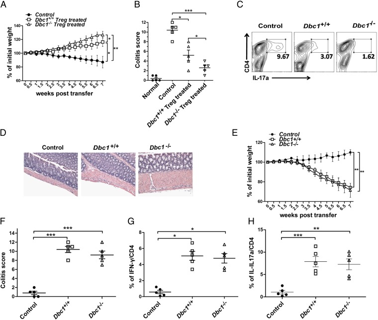
Fig. 4.
Dbc1−/− Treg cells function profoundly in preventing colitis. (A) The changes in weight in the different groups in the colitis model. Treg cells (5 × 10⁵) from the indicated mice were expanded for 4 d and then were cotransferred i.p. with 3 × 10⁵ syngeneic CD4+CD45RBhi T cells into 8- to 10-wk-old Rag2−/− mice (n = 6 mice in each group). Rag2−/− mice receiving only CD4+CD45RBhi T cells were used as a control. Body weights were calculated twice a week. (B) Colitis scores were calculated according to pathological anatomy and histological expression in the different groups. Each data point represents an individual mouse. (C) The percentages of cells expressing IL-17a in the CD4+ population. Fresh cells taken from spleens of mice in the Dbc1−/−, Dbc1+/+, and control groups were stimulated with phorbol12-myristate13-acetate (PMA) and Ionomycin for 1 h and with brefeldin A (BFA) for 4 h (5 h total), followed by staining for IL-17a. (D) Representative photomicrographs of colonic sections from control recipients (n = 11) or recipients that received Dbc1+/+ (n = 7) or Dbc1−/− (n = 3) Treg cells. Data from six independent experiments were pooled. (Original magnification: 200×.) (E) The changes in weight in the different groups in the colitis model. Syngeneic CD4+CD45RBhi T cells (3 × 10⁵) from Dbc1+/+ and Dbc1−/− mice were transplanted i.p. into 8- to 10-wk-old Rag2−/− mice (n = 4 mice in each group). Rag2−/− mice injected with PBS were used as the control group. Body weights were calculated twice a week. (F) Colitis scores were calculated according to the pathological anatomy and histological expression in the different groups. Each data point represents an individual mouse. (G and H) Mesenteric lymph nodes were isolated and stimulated with PMA plus Ionomycin and BFA. Cells were stained for IFN-γ/IL-17a/CD4 and then were analyzed by flow cytometry. *P < 0.05; **P < 0.02; ***P < 0.01.

