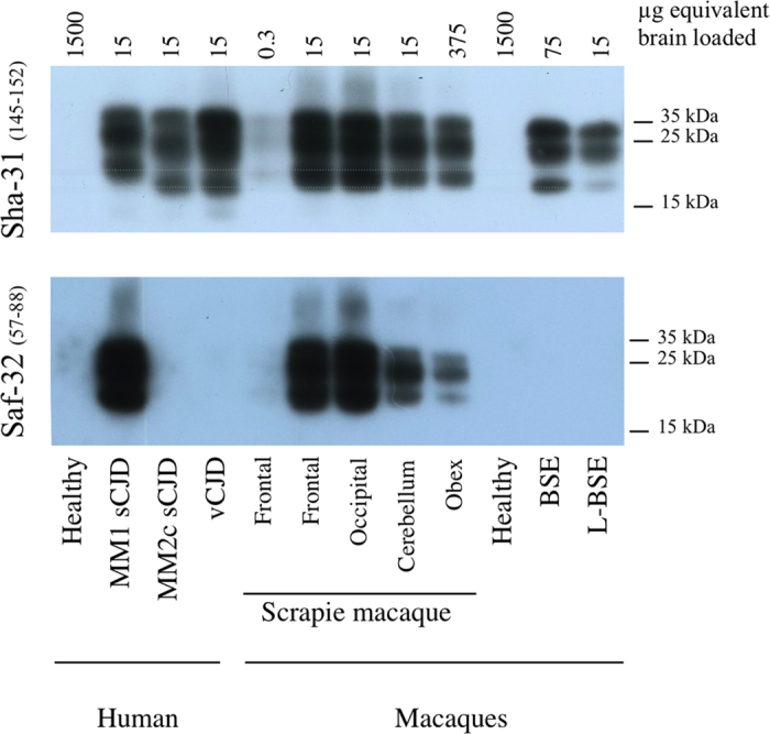
Figure 4. PrPres electrophoretic profile in scrapie-infected macaque.

Samples issued from different brain regions of the scrapie-infected macaque (frontal, occipital, cerebellum or obex) were homogenized (20% weight/volume in 5% glucose solution) and then purified according to the TeSeE purification protocol using 4 μg of proteinase K / mg of brain19. As controls, samples derived from occipital cortices of patients (healthy, MM1 or MM2c sporadic CJD, variant of CJD) or classical BSE- or L-type BSE-infected macaques were equally treated. Resulting purified PrPres was solubilized in loading buffer, the equivalent of 0.3 to 1,500 μg of brain was loaded on a 12% acrylamide gel (depending on the levels of positivity of each sample), and revealed using anti-PrP monoclonal antibodies targeted against the core (Sha-31 recognizing epitope 145–152) or the octapeptides (SAF-32, recognizing octarepeats included in region 57–88) of the protein.
