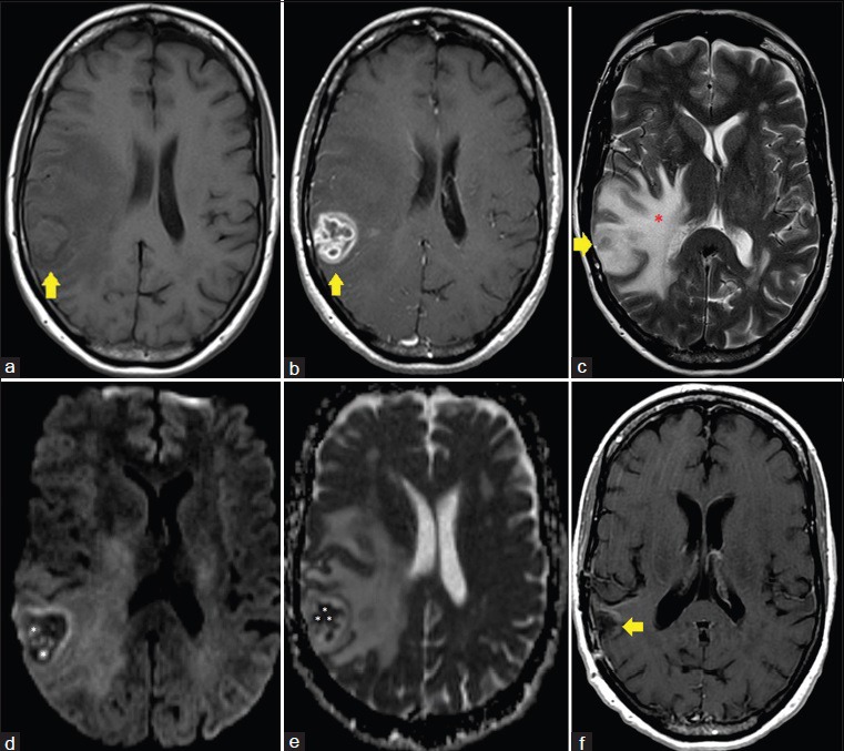
Figure 1.

58-year-old Caucasian female presented with a 6-week history of dizziness upon standing and was diagnosed with cerebral blastomycosis. (a) Axial T1 image, pre-gadolinium administration, shows a 3.0 cm anteroposterior × 2.4 cm transverse × 3.1 cm craniocaudal lesion within the right temporoparietal region (yellow arrow). (b) Axial T1 image, post-gadolinium administration, shows a 3.0 cm anteroposterior × 2.4 cm transverse × 3.1 cm craniocaudal enhancing lesion within the right temporoparietal region (yellow arrow). (c) Axial T2 image demonstrates marked surrounding vasogenic edema throughout the right cerebral hemisphere (red *). (d) Axial diffusion-weighted image demonstrates multiple rounded foci of reduced diffusivity within the central aspect of the lesion (white *). (e) Axial apparent diffusion coefficient (ADC) map image demonstrates multiple rounded foci of reduced diffusivity within the central aspect of the lesion (white *). (f) Axial T1 image, post-gadolinium administration, following resection and antifungal treatment of the lesion demonstrates a resection cavity in the area of the previously seen enhancing lesion with no evidence of residual enhancing infection. The patient is asymptomatic 8 months post resection.
