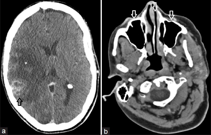
Figure 2.

58-year-old Caucasian female presented with a 6-week history of dizziness upon standing and was diagnosed with cerebral blastomycosis. (a) Axial contrast enhanced CT image of the head obtained intraoperatively demonstrates the rim enhancing lesion within the right temporoparietal region (black arrow) resulting in significant surrounding edema (*). (b) Intraoperative axial contrast enhanced CT image at the level of the paranasal sinuses demonstrates normal aeration of the paranasal sinuses (white arrows) without evidence of mucosal thickening or other abnormality.
