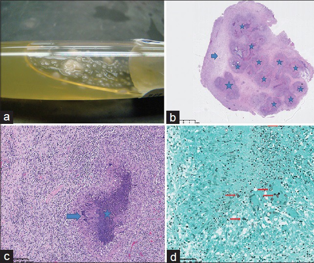
Figure 3.

58-year-old Caucasian female presented with a 6-week history of dizziness upon standing and was diagnosed with cerebral blastomycosis. (a) Photograph of aspirated fluid from the parenchymal abscess demonstrates yeast formation (white droplets) within purulent fluid. (b) Photomicrograph of microscopic examination of the resected lesion: low-power view with 1.5 × magnification shows multiple necrotizing granulomas (stars) and a rim of reactive brain tissue (arrow). The granulomas correspond to areas of decreased signal seen on diffusion-weighted imaging. (c) Photomicrograph of microscopic examination of the resected lesion: high-power view of the granuloma with 40 × magnification shows a rim of palisading histiocytes (arrow) with central necrosis (star). d) Photomicrograph of microscopic examination of the resected lesion. stained with Grocott-methenamine silver (GMS) stain at 40× magnification, highlights the budding yeast of Blastomyces dermatitidis (arrows).
