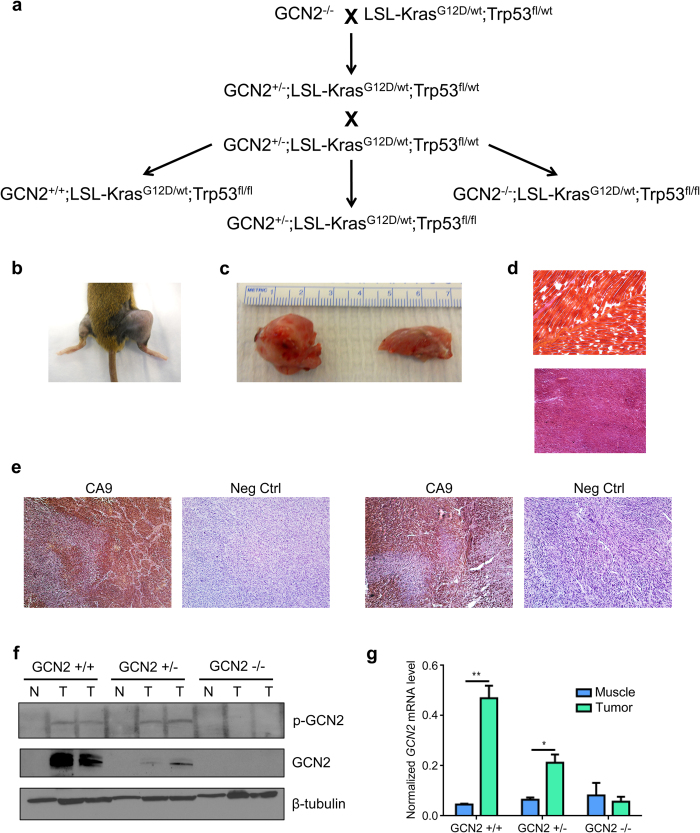
Figure 1. Development of a genetically engineered mouse model to study the role of GCN2 in sarcomagenesis.
(a) Breeding scheme to generate GCN2+/+, GCN2+/−, and GCN2−/− mice on an LSL-KrasG12D/wt;p53fl/fl background. (b) Gross morphology of sarcomas compared to normal leg. The right leg was injected with Ad-cre, while the left leg served as a normal tissue control. (c) Gross morphology of sarcomas compared to normal leg. The size of a typical sarcoma (left) is compared to the size of the normal leg (right) from the same mouse. (d) Typical histology of normal muscle (top) and soft tissue sarcoma (bottom) stained with hematoxylin and eosin. Magnification is 40X. Normal muscle and tumor shown are from the same animal. (e) Typical immunohistochemistry for CA9 in two sarcomas, along with the no primary antibody control. Tissue sections were counterstained with hematoxylin. Magnification is 100X. (f) Western blot analysis of phosphorylated and total GCN2 in homogenized normal muscle tissue (N) and soft tissue sarcomas (T). β-tubulin was used as a loading control. (g) qPCR analysis of GCN2 levels in sarcoma and muscle tissue. GCN2 levels were normalized to the geometric mean of the reference genes β-actin and 18S rRNA. Data are represented as the average value for each genotype ± standard error of the mean; *p < 0.05, **p < 0.01.

