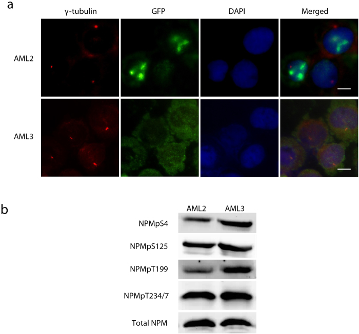
Figure 6. T199 is likewise hyperphosphorylated in NPMc-expressing cell line OCI-AML3.
(a) Immunofluorescence co-staining of centrosome marker γ-tubulin (red puncta) and NPM (green) show that OCI-AML3 (AML3) has cytoplasmic NPM while OCI-AML2 (AML2) has wtNPM which is localised in the nucleus and separated from the centrosome by the nuclear envelope. Bar = 10 μM. (b) Western blot of selected NPM phosphorylation sites. T199 is more phosphorylated in AML3 compared to AML2 whereas S4 phosphorylation is moderately elevated in AML3. Other phosphorylation sites are not significantly different.

