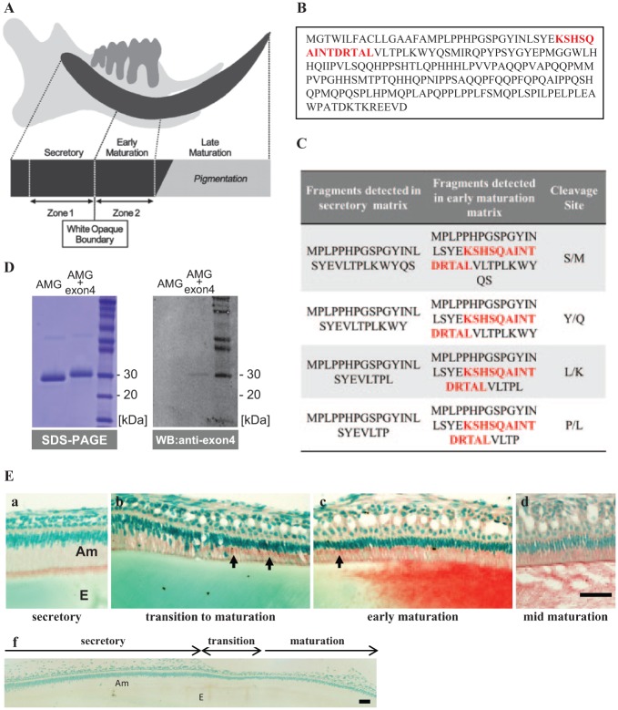
Figure 1.
Identification of amelogenin fragments containing exon4 in enamel matrix. (A) Diagram of a 9-week-old rat incisor showing the location of zone 1, which is primarily a secretory-stage enamel incisor, and zone 2, which is primarily early-maturation-stage enamel. (B) The protein sequence for the full-length amelogenin isoform containing 194 amino acids, with the sequence derived from exon4 shown in red. (C) Protein fragments detected by MS show that exon4 peptide (red font) is only found in the early maturation enamel matrix. (D) Purified RH189 (AMG+4) and RH175 (AMG-4) were separated by SDS PAGE, and then analyzed by Western blot to confirm that the anti-exon4 antibody detected only amelogenin containing exon4 (RH189). (E) Immunodetection of exon4 in incisor ameloblasts showed (a) secretory ameloblasts with positive staining at the apical border, but no obvious staining of the enamel matrix. Beginning in transition and early-maturation-stage ameloblasts (b, c), intense exon4 peptide immunoreaction was seen in the Golgi region (arrows) and in the enamel matrix, consistent with the MS analysis. Pre-immune staining of the incisor shows no nonspecific staining of matrix cells (f). Am, ameloblasts; E, enamel. Bars: 50 μm.

