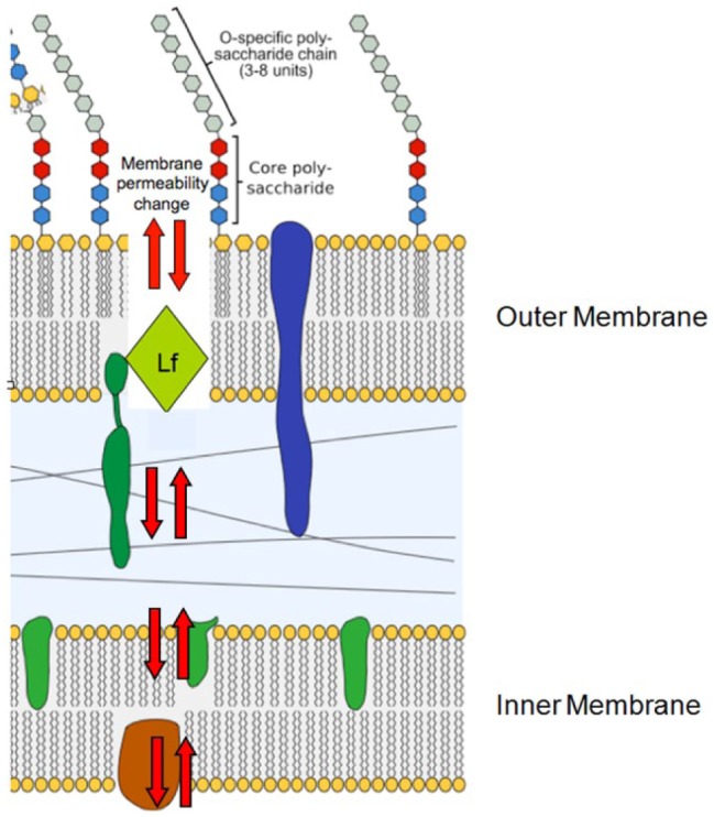
Figure 2.

Illustration of proposed direct effect of lactoferricin on Gram-negative cell wall. Gram-negative cell wall is composed of 3 units: an outer O-specific polysaccharide that determines antigenic specificity, a core polysaccharide, and a lipid A inner structure (also known as endotoxin). Lactoferrin initially depolarizes the membrane by attacking the region where fatty acid tail joins polar head of lipid A. Lipid bilayer is composed of upper region head and tail and a lower region that consists of a tail-to-head configuration. Head is represented as circle or hexagon; fatty acid/hydrophobic tail is represented as squiggly lines beneath head. Lactoferricin insertion between leaflets of the bilayer creates holes in membrane and increased membrane permeability resulting in disturbance of membrane function. Lactoferricin participates in creating physical holes (red arrows) and induces production of hydrolases that degrade the cell wall. This activity causes destabilization of the membrane and leads to redistribution of membrane lipids between leaflets of the bilayer. Lf, lactoferrin.
