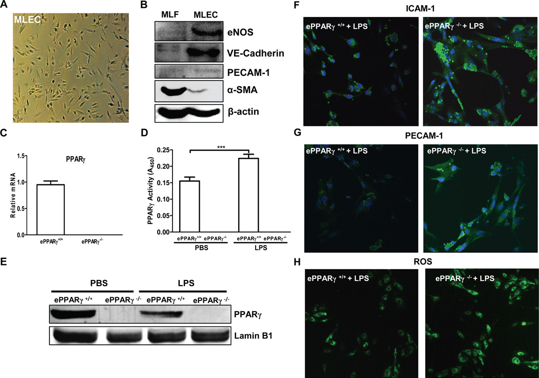
Figure 5. Absence of ePPARγ Exaggerates Inflammatory Responses of MLEC in Vitro.
MLEC were isolated from ePPARγ+/+ and ePPARγ−/− mice (n = 7–8 mice per group) and plated onto 2% gelatin-coated dishes. (A) MLEC exhibit expected endothelial cell morphology. (B) Western blotting showed the expected expression profiles in MLEC and similarly isolated fibroblasts (MLF), respectively, of the endothelium-specific proteins eNOS, vascular endothelial (VE) cadherin, and PECAM-1 and the fibroblast-specific protein α-smooth muscle actin (α-SMA). (C) Relative PPARγ mRNA levels were determined by real-time RT-PCR. In some experiments MLEC were isolated from ePPARγ+/+ and ePPARγ−/− mice 12 h after treatment with LPS (10 mg/kg, i.p.), and nuclear protein extracts prepared. (D) PPARγ DNA-binding activity and (E) protein expression by Western blotting were determined. In other experiments after 2 h serum deprivation, monolayer cultures were treated with LPS (100 ng/ml) for 6 h. Following LPS treatment, immunofluorescence microscopy revealed increased ICAM-1 and PECAM-1 in MLEC isolated from (F,G; right panels) ePPARγ−/− vs. those from (F,G; left panels) ePPARγ+/+ mice, and also elevated (H; right panel vs. left panel) intracellular ROS levels. Blots and images are representative of 3 independent experiments.

