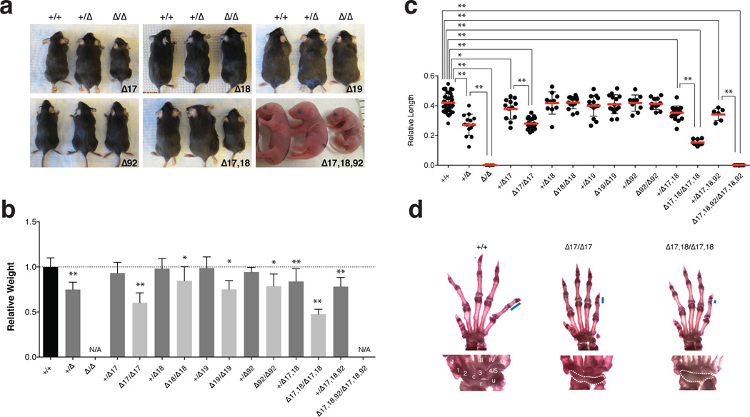
Figure 2. Feingold Syndrome features in the miR-17~92 mutant mice.
(a) Macroscopic appearance of miR-17~92 allelic series’ animals. (b) Mean weight of 4–5 week-old +/+ (n = 77), +/Δ17~92 (n = 13), +/Δ17 (n = 21), Δ17/Δ17 (n = 11), +/Δ18 (n = 26), Δ18/Δ18 (n = 17), +/Δ19 (n = 19), Δ19/Δ19 (n = 6), +/Δ92 (n = 9), Δ92/Δ92 (n = 9), +/Δ17,18 (n = 22), Δ17,18/Δ17,18 (n = 7) and +/Δ17,18,92 (n = 12) animals,– normalized to sex- and age-matched controls. (c) Mean length of the 5th mesophalanx relative to the metacarpal bone. (d) Top, representative pictures of forelimb autopods of wild type, miR-17~92Δ17/Δ17 and miR-17~92Δ17,18/Δ17,18 adult animals. Blue bars represent length of mesophalanx; Bottom, detailed view of wrist bones. Digits are numbered I–V. Carpal bones, 1–3 and 4/5, c (central element), u (ulnare), and r (radiale). Fusion of carpal bones is highlighted with dashed line.
Error bars = s.d.,* p < 0.05, ** p < 0.001, 2-tailed t-test, N/A, not available.

