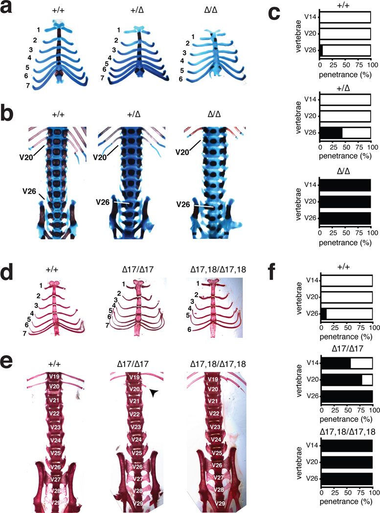
Figure 3. MiR-17~92 regulates axial patterning.
(a) Alcian blue/alizarin red stained rib cages from miR-17~92+/+, miR-17~92+/Δ, and miR-17~92Δ/Δ E18.5 embryos. (b) Ventral view of the axial skeleton. The positions of V20 and V26 are indicated (c) Percentage of animals showing vertebral transformations (black) or normal vertebral identity (white) in miR-17~92+/+ (n = 20), miR-17~92+/Δ (n = 28), and miR-17~92Δ/Δ (n = 10) animals. (d) Alizarin-red stained rib cages from wild type, miR-17~92Δ17/Δ17 and miR-17~92Δ17,18/Δ17,18 adult mice. (e) Ventral view of axial skeleton. Arrow indicates a rudimental rib (f) Percentage of mutant animals showing vertebral transformations (black) or normal vertebral identity (white) in miR-17~92+/+ (n = 97), miR-17~92Δ17/Δ17 (n = 9), and miR-17~92Δ17,18/Δ17,18 (n = 7) animals.

