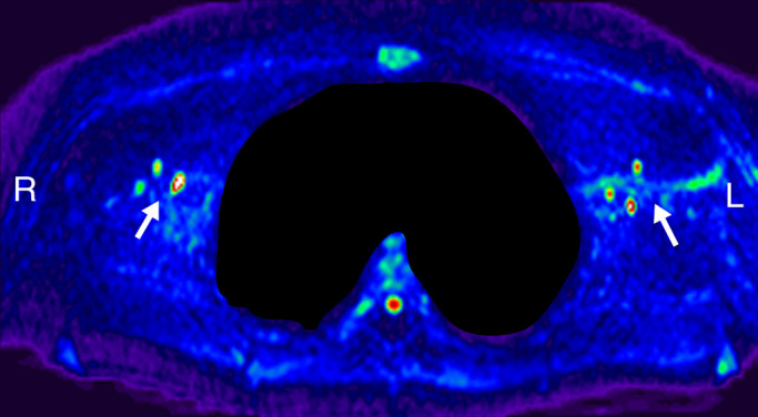
Figure 2c:

Lymph node identification. (a-c) Representative DWIBS MR images in a 30-year-old male patient clearly show the lymph nodes across orthogonal axes within the white rectangles. A typical axial section (c) along the white line in a is used to guide the section location for spin labeling. (d) Corresponding spin labeling MR image in a control subject. Location and planning were guided by DWIBS contrast. (e) DWIBS MR image overlaid on d and thresholded to identify different structures (green = cerebrospinal fluid, yellow = lymph, red = outline of cardiac tissue and major blood vessels) and to draw the regions of interest (two to four voxels) to evaluate lymph kinetic curves.
