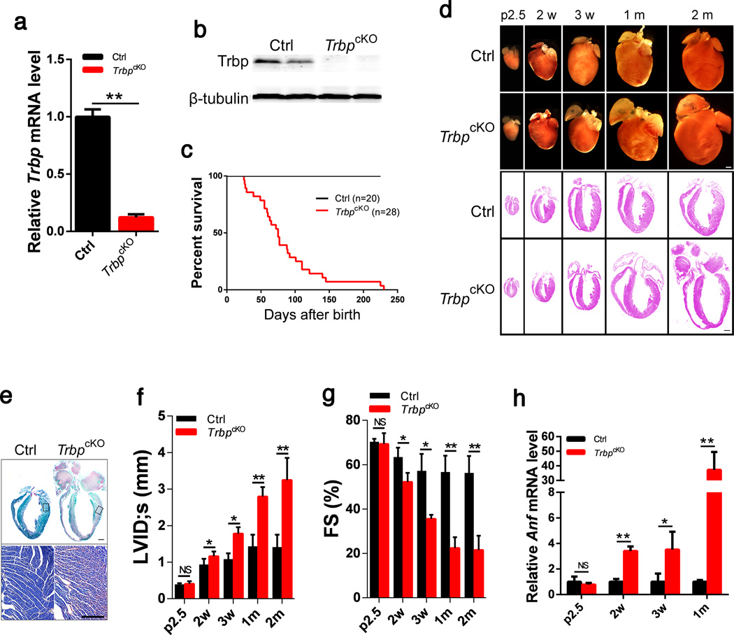
Figure 1. Cardiac-specific Trbp knockout resulted in contraction defects in the heart.
a) qRT-PCR of Trbp mRNA levels in the hearts of postnatal day 2.5 cTNT-CreTG;Trbpfl/fl (TrbpcKO) and control littermate (Ctrl) mice. n = 3.
b) Trbp protein levels in the hearts of postnatal day 2.5 TrbpcKO and control mice were assayed with Western blot. β-tubulin serves as a loading control.
c) Kaplan-Meier survival curves of TrbpcKO and control mice. P<0.01.
d) Gross morphology and histology of TrbpcKO and control hearts at different time points. p2.5: postnatal day 2.5; 2w: 2 weeks after birth; 3w: 3 weeks after birth; 1m: 1 month after birth; 2m: 2 month after birth. Bars = 1.0 mm.
e) Fast green and Sirius red staining of TrbpcKO and control hearts from 2-month old mice. The portion of the heart represented in high magnification is boxed. Bar = 1.0 mm (upper panel); Bar = 500 µm (lower panel).
f) Echocardiography of left ventricle internal dimension at systolic (LVID;s) in TrbpcKO and control mice at indicated time points. n = 3–6.
g) Fractional shortening (FS%) of TrbpcKO and control mice at indicated time points. n = 3–6.
h) Anf mRNA level in the hearts of TrbpcKO and control mice at indicated time points. n = 3. Values are expressed as the mean ± SD. NS: not significant, *: P<0.05, **: P<0.01.

