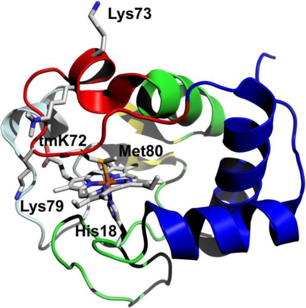
Fig. 1.

Structure of iso-1-Cytc (pdb code: 2YCC, [7]). The loop shown in red is Ω-loop D and provides Met80 as a heme ligand in the native state. Lysines 73 and 79 provide the alkaline state ligands for wild type iso-1-Cytc. Lysine 72 is trimethylated, tmK72, when iso-1-Cytc is expressed in its native host yeast. tmK72 can be seen to lie across the surface of the heme crevice loop, Ω-loop D. The cooperative substructures of Cytc, as defined by Englander and coworkers [13-15], from least to most stable are color-coded: pale cyan, red, yellow, green and blue
