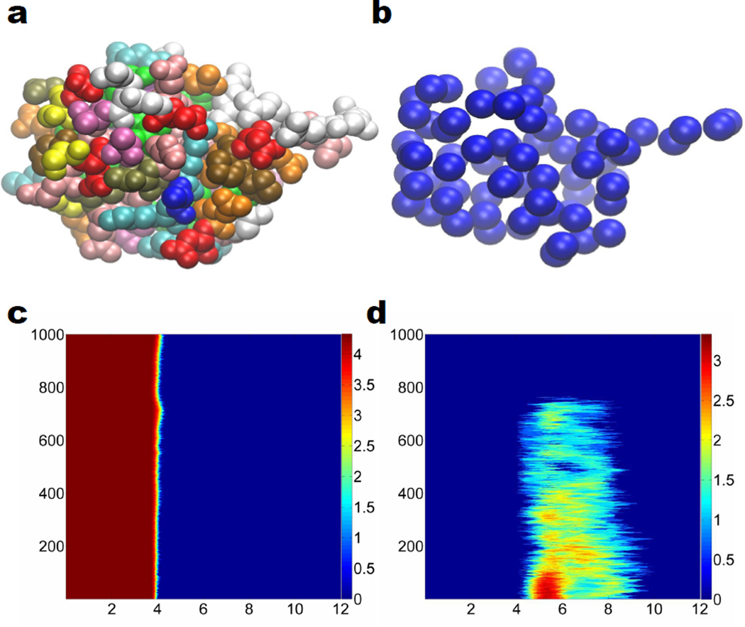
Figure 3.
Coarse-grained representation of the unfolding of protein 1UBQ and the corresponding multidimensional persistence. a All atom representation of the relaxed structure without hydrogen atoms; b Coarse-grain representation of the relaxed structure without hydrogen atoms; c 2D β0 persistence; d 2D β1 persistence. The color in subfigure a denotes different residues. In subfigures c, and d, horizontal axises label the filtration radius (Å) and the vertical axises are the protein configuration index. Color bars denote the natural logarithms of PBNs. We systematically add 1 to all PBNs to avoid the possible logarithm of 0.

