Fig. 1.
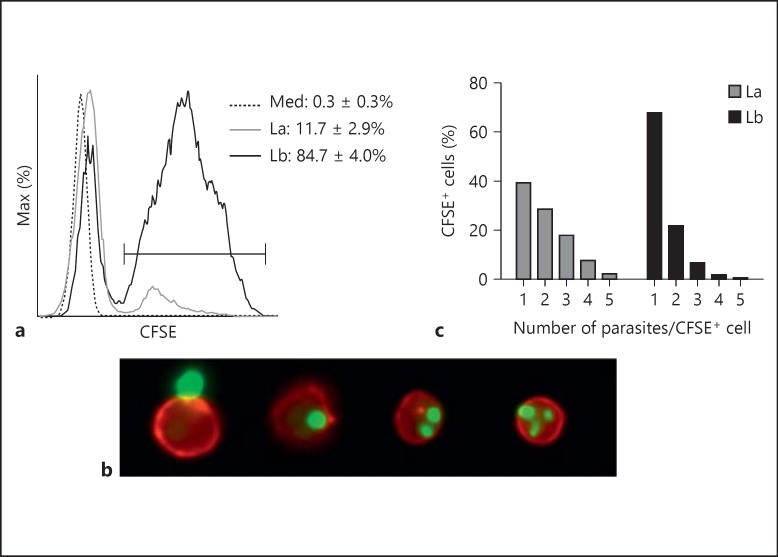
Neutrophil uptake of L. amazonensis and L. braziliensis amastigotes. Thioglycollate-induced peritoneal neutrophils were cocultured with CFSE-labeled amastigotes (MOI of 5) for 4 h. Cells were then analyzed by both FACS (a) and imaging flow cytometry (b, c) to assess parasite uptake. a A histogram shows CD11b+ Ly6G+ neutrophil uptake of CFSE-labeled parasites. b Representative events from imaging flow cytometric analysis showing (left-right) an uninfected neutrophil (red) in close contact with an amastigote (green) and neutrophils infected with 1, 2 and 3 parasites, respectively. c Graphic representation of imaging flow cytometry, showing the percentage of infected cells carrying the designated number of L. amazonensis or L. braziliensis parasites. La = L. amazonensis-infected neutrophils (solid gray line); Lb = L. braziliensis-infected neutrophils (solid black line); Med = uninfected neutrophils (dotted line).

