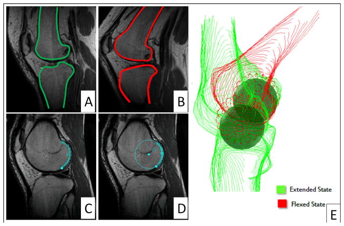
Figure 1.
Representative segmentations of the tibia and femur in (A) extension and in (B) flexion, and examples of (C) manually-defined posterior femoral condyle and (D) automatic segmentation of posterior condyle from previously defined femur segmentation, with (E) three-dimensional reconstruction and modeling of femoral condyles as spheres.

