
Figure 1.
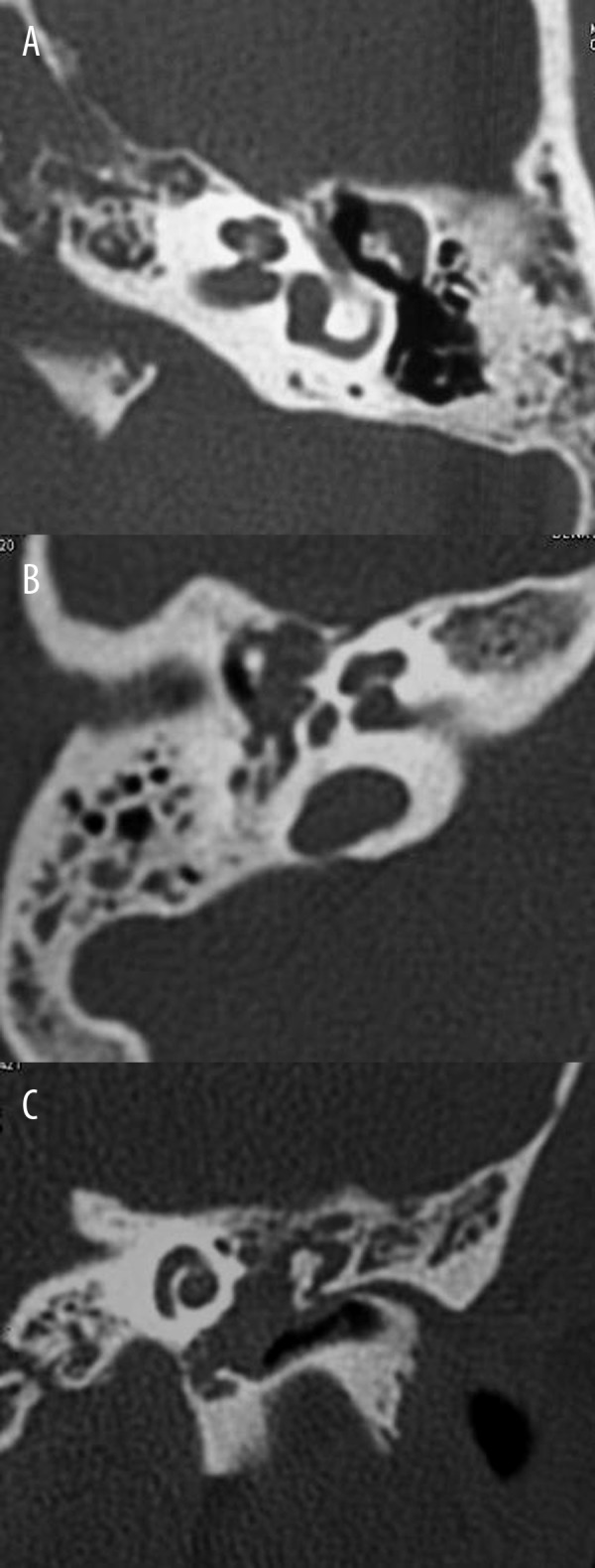
Location of cholesteatoma within the tympanic cavity (T): (A) Axial CT scan of the petrous bone shows attic cholesteatoma located in the attic region lateral to the ossicles (T1). (B) Axial CT scan shows tympanic cholesteatoma located in the tympanic region medial to the ossicles (T2). (C) Coronal CT scan shows atticotympanic cholesteatoma filling the whole middle ear cavity (T3).