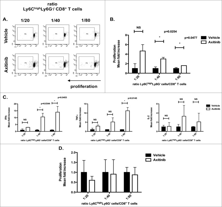
Figure 7.
Axitinib induces differentiation of CD11b+Ly6ChighLy6G– cells to an antigen-presenting phenotype in a subcutaneous, but not in an intracranial tumor model. CD11b+Ly6ChighLy6G– cells were sorted from the subcutaneous and intracranial tumors of MO4-bearing mice on the 7th day of treatment with axitinib at 25 mg/kg bid or vehicle by oral gavage and used in an allogeneic mixed lymphocyte reaction. Controls included T cells cultured in the absence of MDSCs with and without T-cell stimulation. CD11b+Ly6ChighLy6G- cells sorted from subcutaneous tumors of axitinib-treated induced significant more proliferation (B.), and a significantly increased IFNγ, TNFα, and IL2-secretion was found in the supernatant of those proliferating C3H CD8+ T cells as compared to those sorted out of vehicle-treated mice (C.). (A). Representative FACS profile of the proliferation of CD8+ T cells (C3H mouse) in the presence of different ratios of Ly6ChighLy6G- MDSC (moMDSC). (B–C.) Subcutaneous tumor model. (B.) Overview of the percentage of proliferation of the CD8+ T cells (represented as mean fold increase) as induced through co-culture with Ly6ChighLy6G- (moMDSC) in different ratios. (C.) IFNγ, TNFα, and IL-2 production (represented as mean fold increase) by CD8+ T cells was determined after 3 d of co-culture with different ratios of Ly6ChighLy6G- MDSC (moMDSC). Three independent experiments were performed (4 mice per group, tumors were pooled per group before cell sorting) and results are presented as mean ± SEM. (D.) Intracranial tumor model. (D.) Overview of the proliferation of the CD8+ T cells (mean fold increase) as induced through co-cultures with Ly6ChighLy6G- (moMDSC) in different ratios. Two independent experiments were performed (4 mice per group, tumors were pooled per group before cell sorting) and results are presented as mean ± SEM.

