Figure 1.
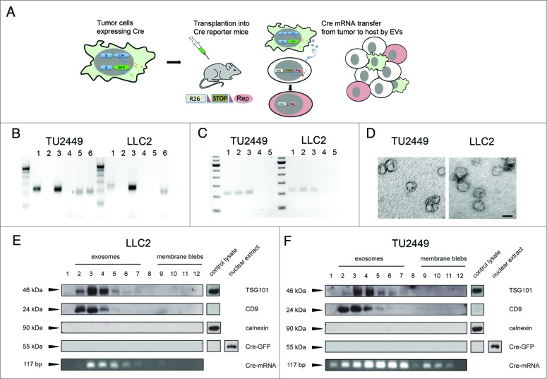
Tumor cells expressing Cre secrete different vesicular subtypes containing Cre mRNA (A) Schematic presentation of the experimental strategy. Tumor cells are stably transduced to constitutively express Cre recombinase and GFP. After transplantation into a Cre reporter mouse, lateral transfer of Cre containing EVs leads to recombination events in the host. (B) Detection of Cre mRNA by RT-PCR in EV preparations from tumor cell conditioned medium. Cre is detectable in the pellet (lane 1) but not in the supernatant (lane 2) after ultracentrifugation. Cre expressing cells served as positive- (lane 3) and omission of reverse transcriptase (RT) from the reverse transcription step as negative controls (lane 4). GFP mRNA was detectable in EVs from glioma- but not carcinoma cells (lane 5), a positive control from GFP expressing cells (lane 6). (C) Cre mRNA is contained in vesicles. Cre mRNA can be detected in EVs (lane 1), also after treatment with Proteinase K (45 min, 2 mg/mL) (lane 2) or Proteinase K followed by RNase digestion (45 min) (lane 3), whereas treatment of EVs with detergent (Triton- X-100, 0.1%) followed by RNase digestion eliminated the signal for Cre (lane 4). RT- negative samples served as negative controls (lane 5). (D) Electron micrographs of vesicle preparations of glioma (TU2449) and carcinoma (LLC2) cell lines showing the cup shaped morphology and size of 50–100 nm typical for exosomes. (F and G) Pelleted (100.000 × g) EVs from cell culture supernatant were loaded on a continuous sucrose gradient and ultracentrifuged. Vesicle subpopulations were tested for all subfractions by blotting against the indicated proteins with cell lysates serving as controls. Fractions 2–7 typically contain exosomes whereas larger vesicles or membrane blebs are contained in fractions 9–12. Cre protein was not detectable in any of the fractions. Representative images of three (B–E) separate sample preparations (Scale bar D, 50 nm).

