
Figure 2.
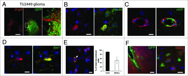
Recombination induced marker gene expression indicates EV uptake (A) Frequent recombination events indicated by immunohistochemical staining for β- galactosidase surrounding the GFP-positive tumor cell mass. Recombined cells included several lineages including neurons (B), endothelial cells (C) and microglia (D). Images are representative from stainings of 2–5 separate animals. (E) Most of recombined cells in the tumor are CD45+ leukocytes (white arrow in left panel) with CD11b+Gr1+ MDSCs as a major subpopulation (right panel). Mean (SD), n = 3. (F) Intracranial injection of EV preparations from Cre expressing glioma cells are sufficient to induce marker gene expression in neural cells (including neurons) in Cre reporter animals (n = 4 injections from 2 separate EV preparations). Scale bars A and E left panel 100 μm, B–E and F right panel, 10 μm.